|
Sediment
geochemistry in action ... lights ... and
cameras....
A
picture is worth a thousand words (or so they say). Below are a number
of sediment geochemistry images, some of which I found while preparing Geochemistry of Marine
Sediments (others I've found since the book was
published).
In retrospect I wish I had included them in the book - I
guess that will have to wait for the second edition (if there ever is
one)
|
|
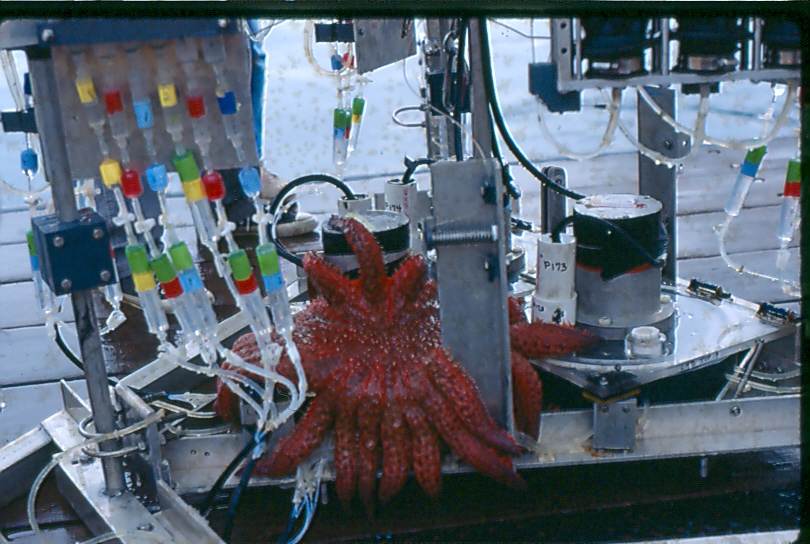
Benthic
ecology meets sediment geochemistry. The USC benthic lander on deck in
Monterey Bay (also see below) with a large starfish attempting to
engulf the oxygen electrode in the lid of the one of the
chambers.
See section 12.1 of the book for a discussion of the use of
benthic
landers to determine sediment-water exchange fluxes.
|
|
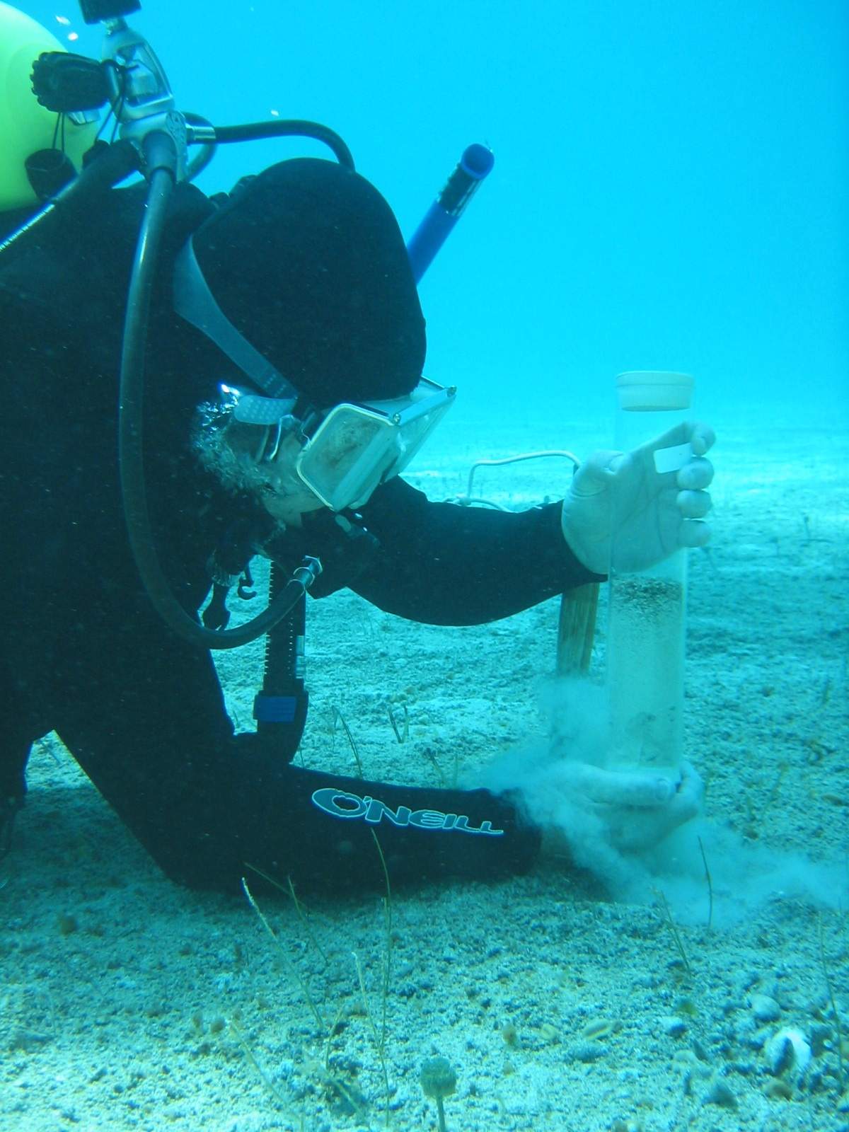
| The
author collecting a short sediment core from shallow water sediments on
the Bahamas Bank. |
|
|
|
|
|
|
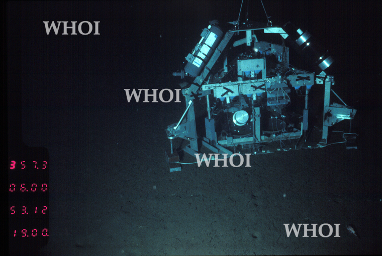
| The USC lander
hard-at-work in Catalina Basin (southern California
Borderland), as photographed from the research submersible Alvin. |
|
|
In situ
photograph of a Mn nodule field in the southwest Pacific Ocean at a
water depth of ~5,000 m. The scale of this photo is roughly 1.5 m x 3
m. See section 13.3 of the book for a discussion of Mn
nodules.
Photo
courtesy of I.C. Wright and can originally be found in New Zealand J. Geol. Geophys.,
2005, vol. 48: 27-41
|
|
|
|
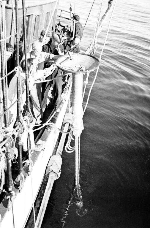
| Collecting a deep-sea
sediment core circa 1930-40 with a Varney-Redwine deep sea corer (image
from the SIO Archives) |
|
 |
|
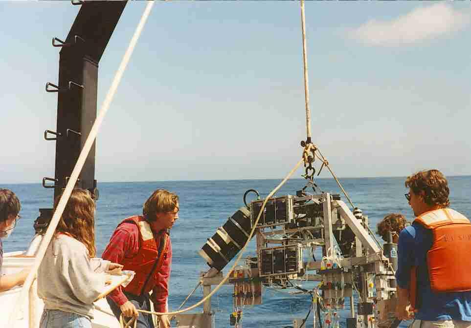
Deploying
the USC benthic lander at sea.
|
|
|
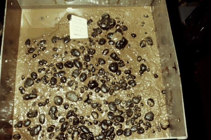
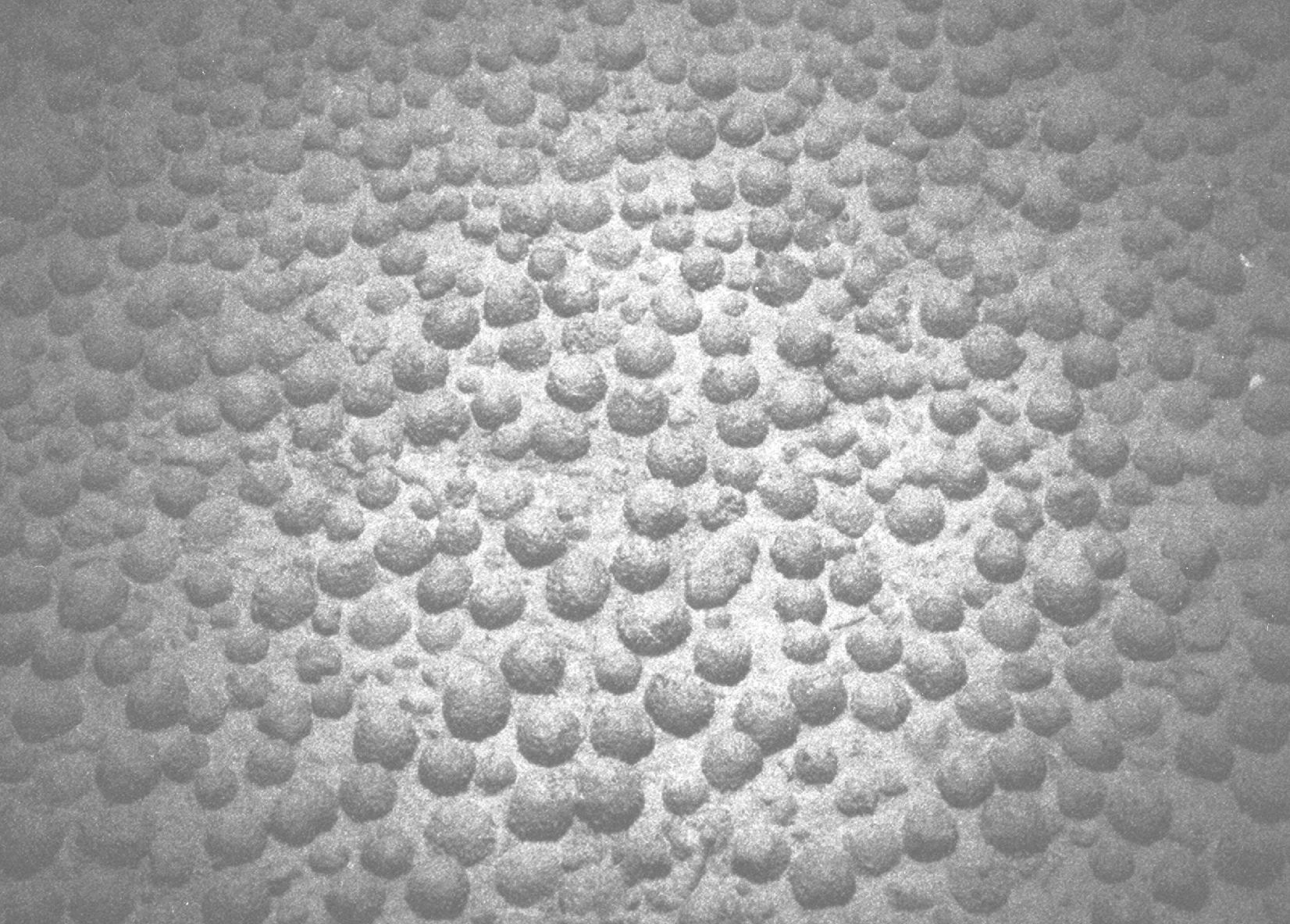
A
box core showing a high density of manganese nodules (section 13.3)
from a site in the tropical Pacific (image is from the NOAA
Photo Library).
|
|
|
|
|
|
|
|
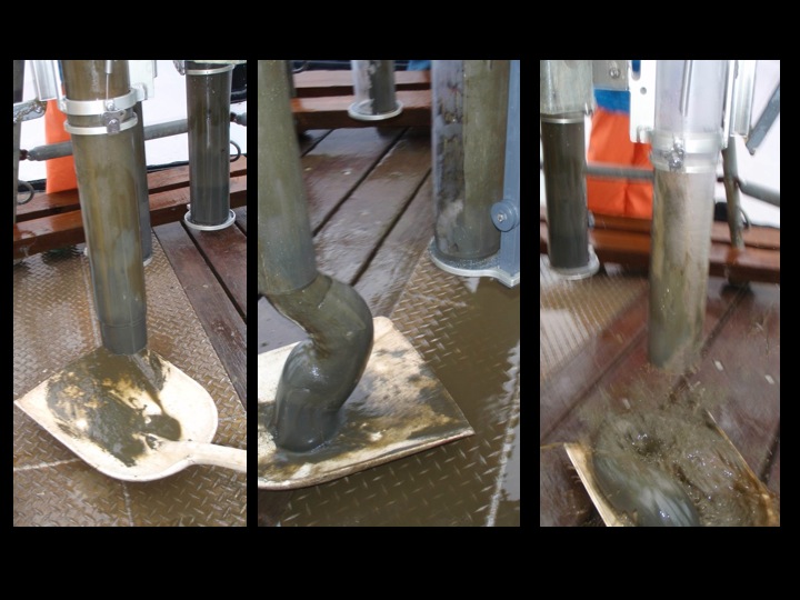
| Cleaning out an
unused sediment core from a multi-core tube (photos courtesy of the late Nuria
Protopopescu) |
|
|
|
|
|
|
|
|
|
|
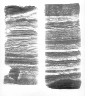
| Photograph
and X-radiograph of a multi-core from Santa Barbara Basin showing
laminated sediments from the center of the basin (see the
Appendix of the book for further
details about Santa Barbara Basin).
Photos courtesy of Lowell Stott, USC. |
|
|
|
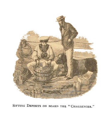
| Early
sediment sampling aboard the HMS Challenger (1872-76) |
|
|
|
|
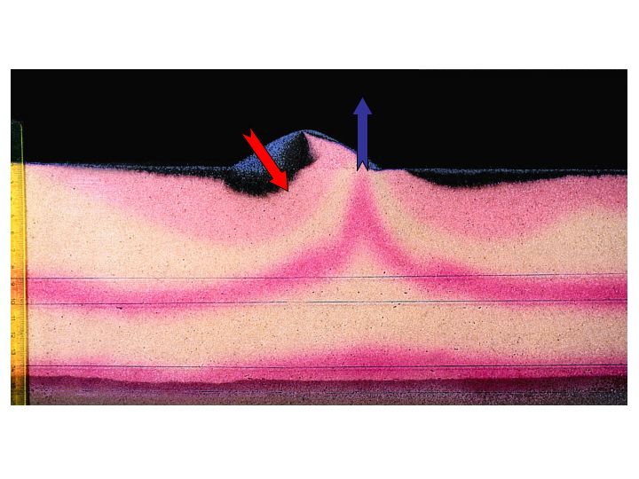
| Flume
studies illustrating the upwelling of red pore fluid (blue arrow) and
intrusion of tracer from the water column (red arrow) caused by
boundary flow-topography interactions in permeable sands (see section
12.3 of the book). Photo courtesy of Marcus
Huettel, Florida State Univ. and can be originally found in Limnol. Oceanogr.,
1996, 41: 309-322. |
|
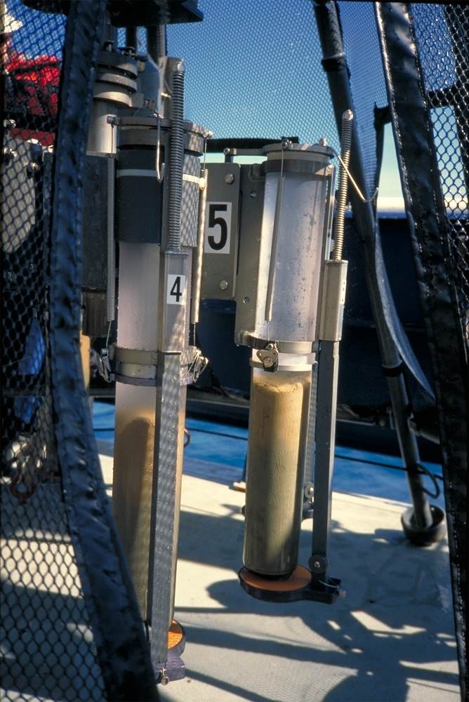
| Sediment
cores collected with a multi-corer from
a site in the eastern tropical Pacific. Note the transition about
halfway down the core from a brown, oxidized layer surface layer to
(reduced ?) greenish-gray clay below (see sections 7.3.3.2 and 13.4 of
the book).
Photo credit: David M. Anderson, NOAA
Paleoclimatology Program. |
|
|
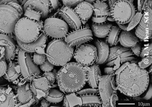
| The
siliceous frustules of the diatoms Skeletonema costatum
(above) andThalassiosira pacifica (below) from
Holocene laminated sediments collected in Saanich Inlet, British
Columbia during ODP Leg 169S (see sections 2.2 and 13.4 of the book).
Source: J.
Dean, SOC. |
|
 |
|
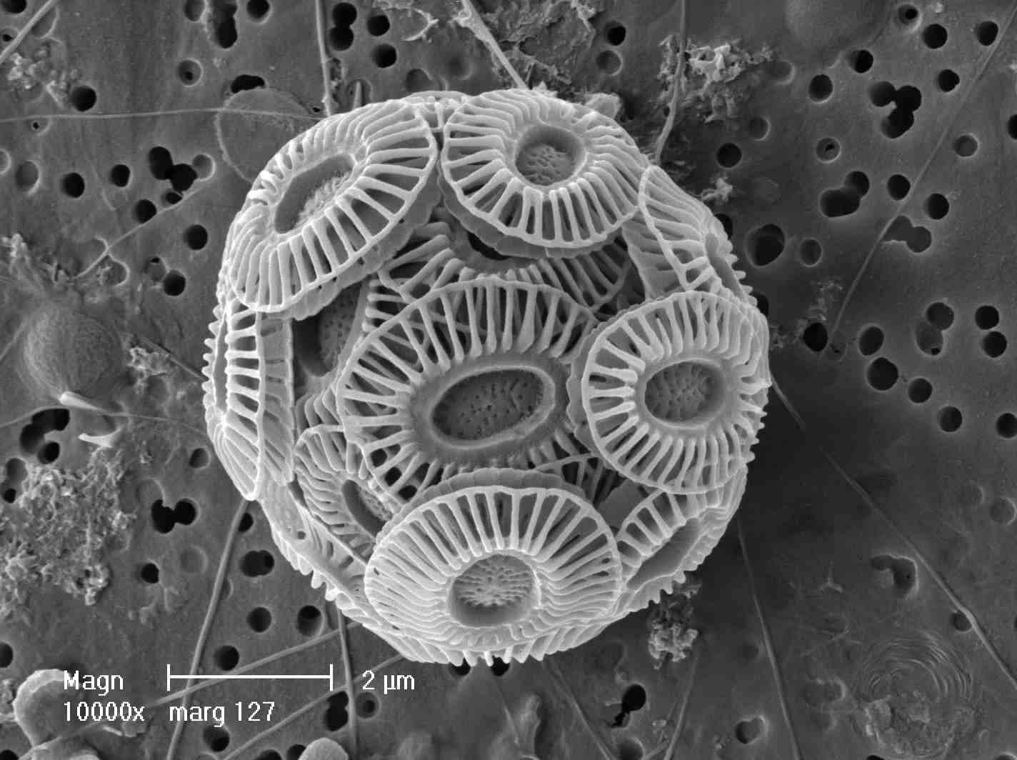
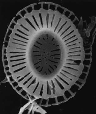
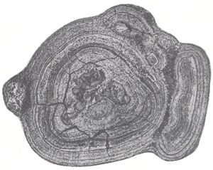
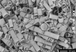
The
coccolithophore Emiliania
huxlyei covered by calcium carbonate platelets (or
coccoliths). An individual coccolith platelet is shown to the right
(see sections 2.2 and 13.5). Source: Toby
Tyrell, SOC.
|
|
|
|
|
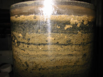
Seasonal
redox zonation in Cape Lookout Bight
sediments. These winter sediments show a several cm "brown"
oxidized layer at the sediment surface, below which the sediments are
anoxic and sulfidic (see section 14.3 of the book).
|
|